The Cellular Factory Floor — ER, Golgi, Lysosomes
A guided tour of the production lines, packaging stations, and recycling crews inside every eukaryotic cell

Imagine a factory that operates around the clock with no electricity bills, no managers, no human workers. It produces thousands of different goods, packages each one for a specific customer, ships them across the building, recycles every scrap of waste, and keeps the entire enterprise running on chemical signals alone.
Such a factory exists. About 30 trillion of them exist inside you, right now. Each one is smaller than a speck of dust.
What kind of arrangement could possibly do all this in a space so small?
Think about what a real factory needs: production lines, packaging stations, transport, waste disposal. Could nature have evolved miniature versions of each?
The Verse on the Cycle of Cooperative Action
अन्नाद्भवन्ति भूतानि पर्जन्यादन्नसम्भवः।
यज्ञाद्भवति पर्जन्यो यज्ञः कर्मसमुद्भवः॥
'अन्न से सारे जीव बनते हैं। अन्न बनता है बारिश से। बारिश आती है यज्ञ (मिल-जुलकर किए गए काम) से। और यज्ञ खुद कर्म (कर्म, सेवा) से होता है।'
"From food, beings arise. From rain, food. From yajña (cooperative offering), rain comes. And yajña arises from action."
The verse names a deep truth: nothing in the universe sustains itself alone. Every part gives to other parts, and receives from other parts. This is yajña — the cycle of cooperative giving. The cell is exactly such a system: ribosomes give proteins to the ER; the ER hands work to the Golgi; the Golgi packages and ships; lysosomes recycle waste back for reuse. Each organelle gives. Each receives. The cell stays alive only because of the giving.
Ribosomes — The Tiny Protein Factories
Begin with the smallest workers. Ribosomes are tiny structures — far too small to see under a light microscope, only visible under an electron microscope. There may be millions of them in a single cell.
Despite their size, ribosomes do one of the most important jobs in all of biology: they make proteins.
Every cell needs proteins, constantly. Some proteins are structural — they hold the cell together. Others are enzymes that drive the cell's chemical reactions. Others are receptors, channels, signals, defenders. Without a steady supply of proteins, no cell can function — let alone divide, repair, or grow.
Ribosomes read instructions from the cell's DNA (transmitted through a related molecule called messenger RNA) and translate those instructions into chains of amino acids — proteins.
Ribosomes can be found in two places in the cell:
- Free in the cytoplasm — making proteins that will work inside the cell.
- Attached to the surface of the endoplasmic reticulum — making proteins that will be exported, or used in cell membranes.
This division of labour leads us directly to the next organelle.
The Endoplasmic Reticulum — The Manufacturing Network
The Endoplasmic Reticulum (ER for short) is a vast network of folded membranes that spreads through the cytoplasm of the cell. The word reticulum literally means network — and that is exactly what it looks like under an electron microscope: a maze of interconnected channels and flattened sacs.
The ER is continuous with the outer membrane of the nucleus — it physically connects to the nuclear envelope. So messages and materials travel naturally from the nucleus into the ER and on into the rest of the cell.
There are two types of ER, and they look distinctly different:
Rough Endoplasmic Reticulum (RER). The RER appears rough under the electron microscope because its surface is studded with ribosomes. Those ribosomes make proteins, and the freshly made proteins are immediately fed into the channels of the RER for processing and transport. The RER is especially abundant in cells that produce a lot of protein for export — for example, the cells of the pancreas that secrete digestive enzymes, or the cells that produce antibodies for your immune system.
Smooth Endoplasmic Reticulum (SER). The SER has no ribosomes attached — its membranes are smooth. Instead of protein synthesis, the SER specialises in:
- Synthesising fats (lipids) — including the lipids that make up cell membranes.
- Synthesising hormones — for example, the steroid hormones produced in your reproductive organs.
- Detoxifying harmful substances in liver cells — breaking down poisons before they can damage the body.
The ER is, in short, the cell's manufacturing department. Both branches together build proteins, fats, and hormones — and ship them on to the next stage in the production line.
The Golgi Apparatus — Packaging and Shipping
Once the ER has manufactured proteins or lipids, those goods don't simply sit there. They have to be finished, sorted, packaged, and shipped — exactly like products in a real factory.
The Golgi apparatus does this job. Look closely at any electron-microscope image of a cell and you will see, near the ER, a stack of flattened, curved, sac-like structures — like a pile of pita breads. That stack is the Golgi apparatus.
Think of the Golgi as the cell's post office. It is functionally connected to the ER, the cell membrane, and other organelles. Its job is fourfold:
- Modify: chemically tweak the proteins and lipids it receives from the ER — adding sugar groups, refining the structure.
- Sort: identify what each molecule is for, and route it to the right destination.
- Package: enclose each finished product in a small membrane-bubble called a vesicle.
- Ship: send the vesicles to where they are needed — to the cell membrane (for export), to lysosomes (for the recycling crew), or back to the ER.
Without the Golgi, the proteins made by the ER would be raw, unaddressed, and undeliverable. The Golgi turns the cell's manufacturing into a coherent supply chain.
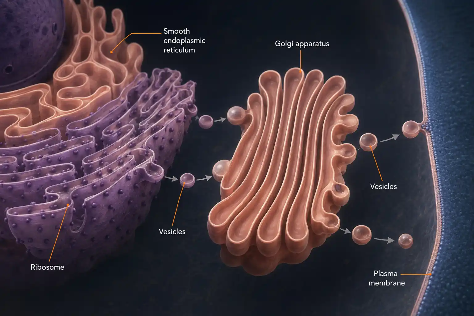
Lysosomes — The Clean-Up Crew
Every busy factory produces waste — broken tools, expired materials, defective products. So does every busy cell. Used proteins. Worn-out organelles. Damaged molecules. Foreign material that has entered the cell. What happens to all of this?
If the waste piled up, the cell would suffocate in its own debris. To prevent that, the cell has a dedicated cleanup system: lysosomes.
A lysosome is a small, single membrane-bound sac filled with digestive enzymes. The enzymes are powerful — they can break down almost any biological molecule: proteins, carbohydrates, fats, nucleic acids, even damaged organelles.
When something needs to be recycled, it is delivered to a lysosome. The lysosome's enzymes break it down into its smallest parts (amino acids, simple sugars, fatty acids). These products are then released back into the cytoplasm, where the cell can reuse them to build new structures.
A crucial design feature: the lysosome's enzymes are only active inside the lysosome's strongly acidic interior. If they leaked into the rest of the cell, they would dissolve the cell from inside out. The lysosome's membrane keeps these dangerous enzymes safely contained — letting them do their work only in their proper place.
This is why lysosomes are sometimes called the suicide bags of the cell. Under certain conditions (for example, when a cell is damaged beyond repair), lysosomes can rupture deliberately, releasing their enzymes and causing the cell to digest itself — a controlled cellular self-destruction. This is one of the body's normal ways of disposing of cells that are no longer needed.
A Surprising Use for Lysosomal Enzymes
Lysosomes are usually thought of as the cell's recycling crew — internal clean-up. But human sperm cells put lysosomal enzymes to a completely different use.
Cells of the pancreas that produce digestive enzymes (these enzymes are exported into your small intestine to digest your food) have an extraordinarily large and well-developed rough endoplasmic reticulum and Golgi apparatus — much more than most other cells.
In contrast, liver cells, which detoxify alcohol and other harmful substances, have an extraordinarily large and well-developed smooth endoplasmic reticulum — but a comparatively modest rough ER.
Why do these two cell types have such different organelle 'profiles'?
Manana Moment
Contemplation before you continue
The Bhagavad Gita's image of yajña — the cycle of cooperative offering — is unusually concrete in this chapter. The ribosome offers protein to the rough ER. The rough ER passes work to the Golgi. The Golgi packages and ships. The lysosome receives waste and returns recycled materials to the cytoplasm. None of these organelles can survive without the others. Each is doing yajña — giving its work to the next station — every moment.
Before you continue, ask yourself:
In your own daily life, where are you receiving work from someone (the food someone cooked, the lessons someone teaches, the cleaning someone did) — and where are you giving your work to others (a homework helped, a chore done, a kindness offered)?
The cell teaches us a startling truth: nothing alive lives alone. Even at the level of organelles inside a single cell, life is a network of small offerings, each one received and passed forward. You are part of countless such cycles — at the cellular, family, community, and ecological level — most of which you have never consciously noticed.
What This Page Teaches Us
-
Ribosomes are tiny structures (free in cytoplasm or attached to the ER) that make proteins by reading instructions from the cell's DNA.
-
The endoplasmic reticulum (ER) is a folded network of membranes throughout the cytoplasm, continuous with the outer nuclear envelope. Two kinds:
- Rough ER has ribosomes attached; specialises in protein synthesis and export.
- Smooth ER has no ribosomes; specialises in fat synthesis, hormone synthesis, and detoxification.
-
The Golgi apparatus is a stack of flattened, sac-like structures that modifies, sorts, packages, and ships proteins and lipids into vesicles for delivery. It is the cell's post office.
-
Lysosomes are single-membrane sacs filled with digestive enzymes. They break down unwanted proteins, carbohydrates, fats, and damaged organelles — keeping the cell clean. The enzymes work only in the lysosome's strongly acidic interior, safely contained.
-
Camillo Golgi discovered the Golgi apparatus in 1898 in barn owl nerve cells, using a silver-staining technique he himself invented. He won the 1906 Nobel Prize.
-
The structure of a cell reflects its function: pancreatic cells (which export proteins) have huge amounts of rough ER and Golgi; liver cells (which detoxify) have huge amounts of smooth ER.
-
Every step of this work depends on cooperation between organelles. The Bhagavad Gita's vision of yajña — the cycle of cooperative offering — is unusually literal at the cellular level: nothing in the cell, and nothing alive on Earth, lives entirely on its own.
Q1.What is the function of ribosomes in the cell?
Imagine a factory that operates around the clock with no electricity bills, no managers, no human workers. It produces thousands of different goods, packages each one for a specific customer, ships them across the building, recycles every scrap of waste, and keeps the entire enterprise running on chemical signals alone.
Such a factory exists. About 30 trillion of them exist inside you, right now. Each one is smaller than a speck of dust.
What kind of arrangement could possibly do all this in a space so small?
Think about what a real factory needs: production lines, packaging stations, transport, waste disposal. Could nature have evolved miniature versions of each?
The Verse on the Cycle of Cooperative Action
अन्नाद्भवन्ति भूतानि पर्जन्यादन्नसम्भवः।
यज्ञाद्भवति पर्जन्यो यज्ञः कर्मसमुद्भवः॥
'अन्न से सारे जीव बनते हैं। अन्न बनता है बारिश से। बारिश आती है यज्ञ (मिल-जुलकर किए गए काम) से। और यज्ञ खुद कर्म (कर्म, सेवा) से होता है।'
"From food, beings arise. From rain, food. From yajña (cooperative offering), rain comes. And yajña arises from action."
The verse names a deep truth: nothing in the universe sustains itself alone. Every part gives to other parts, and receives from other parts. This is yajña — the cycle of cooperative giving. The cell is exactly such a system: ribosomes give proteins to the ER; the ER hands work to the Golgi; the Golgi packages and ships; lysosomes recycle waste back for reuse. Each organelle gives. Each receives. The cell stays alive only because of the giving.
Ribosomes — The Tiny Protein Factories
Begin with the smallest workers. Ribosomes are tiny structures — far too small to see under a light microscope, only visible under an electron microscope. There may be millions of them in a single cell.
Despite their size, ribosomes do one of the most important jobs in all of biology: they make proteins.
Every cell needs proteins, constantly. Some proteins are structural — they hold the cell together. Others are enzymes that drive the cell's chemical reactions. Others are receptors, channels, signals, defenders. Without a steady supply of proteins, no cell can function — let alone divide, repair, or grow.
Ribosomes read instructions from the cell's DNA (transmitted through a related molecule called messenger RNA) and translate those instructions into chains of amino acids — proteins.
Ribosomes can be found in two places in the cell:
- Free in the cytoplasm — making proteins that will work inside the cell.
- Attached to the surface of the endoplasmic reticulum — making proteins that will be exported, or used in cell membranes.
This division of labour leads us directly to the next organelle.
The Endoplasmic Reticulum — The Manufacturing Network
The Endoplasmic Reticulum (ER for short) is a vast network of folded membranes that spreads through the cytoplasm of the cell. The word reticulum literally means network — and that is exactly what it looks like under an electron microscope: a maze of interconnected channels and flattened sacs.
The ER is continuous with the outer membrane of the nucleus — it physically connects to the nuclear envelope. So messages and materials travel naturally from the nucleus into the ER and on into the rest of the cell.
There are two types of ER, and they look distinctly different:
Rough Endoplasmic Reticulum (RER). The RER appears rough under the electron microscope because its surface is studded with ribosomes. Those ribosomes make proteins, and the freshly made proteins are immediately fed into the channels of the RER for processing and transport. The RER is especially abundant in cells that produce a lot of protein for export — for example, the cells of the pancreas that secrete digestive enzymes, or the cells that produce antibodies for your immune system.
Smooth Endoplasmic Reticulum (SER). The SER has no ribosomes attached — its membranes are smooth. Instead of protein synthesis, the SER specialises in:
- Synthesising fats (lipids) — including the lipids that make up cell membranes.
- Synthesising hormones — for example, the steroid hormones produced in your reproductive organs.
- Detoxifying harmful substances in liver cells — breaking down poisons before they can damage the body.
The ER is, in short, the cell's manufacturing department. Both branches together build proteins, fats, and hormones — and ship them on to the next stage in the production line.
The Golgi Apparatus — Packaging and Shipping
Once the ER has manufactured proteins or lipids, those goods don't simply sit there. They have to be finished, sorted, packaged, and shipped — exactly like products in a real factory.
The Golgi apparatus does this job. Look closely at any electron-microscope image of a cell and you will see, near the ER, a stack of flattened, curved, sac-like structures — like a pile of pita breads. That stack is the Golgi apparatus.
Think of the Golgi as the cell's post office. It is functionally connected to the ER, the cell membrane, and other organelles. Its job is fourfold:
- Modify: chemically tweak the proteins and lipids it receives from the ER — adding sugar groups, refining the structure.
- Sort: identify what each molecule is for, and route it to the right destination.
- Package: enclose each finished product in a small membrane-bubble called a vesicle.
- Ship: send the vesicles to where they are needed — to the cell membrane (for export), to lysosomes (for the recycling crew), or back to the ER.
Without the Golgi, the proteins made by the ER would be raw, unaddressed, and undeliverable. The Golgi turns the cell's manufacturing into a coherent supply chain.
Lysosomes — The Clean-Up Crew
Every busy factory produces waste — broken tools, expired materials, defective products. So does every busy cell. Used proteins. Worn-out organelles. Damaged molecules. Foreign material that has entered the cell. What happens to all of this?
If the waste piled up, the cell would suffocate in its own debris. To prevent that, the cell has a dedicated cleanup system: lysosomes.
A lysosome is a small, single membrane-bound sac filled with digestive enzymes. The enzymes are powerful — they can break down almost any biological molecule: proteins, carbohydrates, fats, nucleic acids, even damaged organelles.
When something needs to be recycled, it is delivered to a lysosome. The lysosome's enzymes break it down into its smallest parts (amino acids, simple sugars, fatty acids). These products are then released back into the cytoplasm, where the cell can reuse them to build new structures.
A crucial design feature: the lysosome's enzymes are only active inside the lysosome's strongly acidic interior. If they leaked into the rest of the cell, they would dissolve the cell from inside out. The lysosome's membrane keeps these dangerous enzymes safely contained — letting them do their work only in their proper place.
This is why lysosomes are sometimes called the suicide bags of the cell. Under certain conditions (for example, when a cell is damaged beyond repair), lysosomes can rupture deliberately, releasing their enzymes and causing the cell to digest itself — a controlled cellular self-destruction. This is one of the body's normal ways of disposing of cells that are no longer needed.
A Surprising Use for Lysosomal Enzymes
Lysosomes are usually thought of as the cell's recycling crew — internal clean-up. But human sperm cells put lysosomal enzymes to a completely different use.
Cells of the pancreas that produce digestive enzymes (these enzymes are exported into your small intestine to digest your food) have an extraordinarily large and well-developed rough endoplasmic reticulum and Golgi apparatus — much more than most other cells.
In contrast, liver cells, which detoxify alcohol and other harmful substances, have an extraordinarily large and well-developed smooth endoplasmic reticulum — but a comparatively modest rough ER.
Why do these two cell types have such different organelle 'profiles'?
What This Page Teaches Us
-
Ribosomes are tiny structures (free in cytoplasm or attached to the ER) that make proteins by reading instructions from the cell's DNA.
-
The endoplasmic reticulum (ER) is a folded network of membranes throughout the cytoplasm, continuous with the outer nuclear envelope. Two kinds:
- Rough ER has ribosomes attached; specialises in protein synthesis and export.
- Smooth ER has no ribosomes; specialises in fat synthesis, hormone synthesis, and detoxification.
-
The Golgi apparatus is a stack of flattened, sac-like structures that modifies, sorts, packages, and ships proteins and lipids into vesicles for delivery. It is the cell's post office.
-
Lysosomes are single-membrane sacs filled with digestive enzymes. They break down unwanted proteins, carbohydrates, fats, and damaged organelles — keeping the cell clean. The enzymes work only in the lysosome's strongly acidic interior, safely contained.
-
Camillo Golgi discovered the Golgi apparatus in 1898 in barn owl nerve cells, using a silver-staining technique he himself invented. He won the 1906 Nobel Prize.
-
The structure of a cell reflects its function: pancreatic cells (which export proteins) have huge amounts of rough ER and Golgi; liver cells (which detoxify) have huge amounts of smooth ER.
-
Every step of this work depends on cooperation between organelles. The Bhagavad Gita's vision of yajña — the cycle of cooperative offering — is unusually literal at the cellular level: nothing in the cell, and nothing alive on Earth, lives entirely on its own.
Q1.What is the function of ribosomes in the cell?